Tsetse Flies
Glossina morsitans morsitans & Glossina palpalis

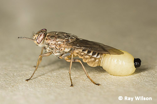
bloodfed female Savannah Tsetse fly (Glossina morsitans morsitans)
Tsetse flies are obligate haematophagous insects, meaning both male and female flies survive purely on a diet of blood. Through their bite, infected flies are capable of transmitting the single-celled protozoan parasite Trypanosoma brucei which causes African Sleeping Sickness (or African Trypanosomiasis) in humans and Nagana in cattle. Through a program of control and surveillance the incidence of human trypanosomiasis has dropped from a peak of nearly 500000 new cases in 1997 to the current level of about 50000 new cases of trypanosomiasis per year in the 36 countries of sub-saharan Africa where the Tsetse fly resides. The economic cost in lost revenue to the African continent due to the effects of nagana on livestock is estimated at US$4.5billion per year.

bloodfed female Savannah Tsetse fly (Glossina morsitans morsitans)
Early symptoms of the disease are similar to flu (fever, joint pains and headaches), but these progress to severe swelling of the lymph nodes as the parasites multiply in the blood. If left untreated, the disease will eventually progress to produce the neurological disorders that give the disease its name, with patients having disrupted sleep patterns, reduced coordination and becoming confused. Once it reaches this stage the neurological damage is often irreversible and if allowed to continue its progression is almost always fatal.
The Tsetse Fly Life-cycle
The Tsetse fly has a quite unusual life-cycle for an insect. Most invertebrates have a reproductive strategy that involves producing large numbers of offspring to offset losses through predation and to enable rapid increases in population whenever possible. Tsetse flies, however, only produce one larva at a time and incubate each within their bodies until the larva is fully developed. Within its lifetime, each female is only capable of raising a handful of offspring.
1. Courtship & Mating
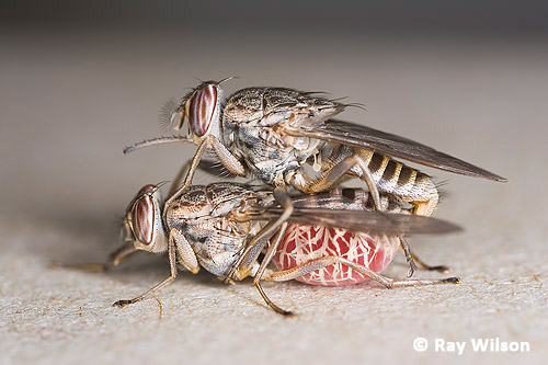
Copulating Savannah Tsetse Flies (Glossina morsitans morsitans)
The female has a recent bloodmeal filling her abdomen
Females are receptive to mating almost immediately after emergence from their puparium. In the wild, mating probably occurs close to, or even on, the host animal around the time of the female's first bloodmeal. There is no courtship involved, with the male simply landing on the female's back. Copulation can last for 1-2 hours, during which the male deposits a ball of sperm at the entrance to the spermathecal ducts. From there the sperm swim up towards the spermatheca, where they remain in a viable state for the remainder of the female's life. The female does not need to mate again.
2. Pregnancy
About 7-9 days after emergence a single fertilized egg is implanted in the female's uterus. The embryo develops inside the female's body, hatches, and the larva completes its full growth cycle in the uterus.
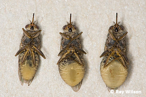
Stages in the pregnancy of female Savannah Tsetse fly (Glossina morsitans morsitans) showing the growth of the larva within her.
Whilst within the abdominal cavity, the larva is fed on a secretion from a specialised milk gland which is deposited close to the head of the larva so that it can directly suck the milk into its gut. The larva breathes through its abdominal polypneustic lobes and gets its oxygen supply directly from air passing in through the female's vulva.
The larva undergoes two moults whilst within the female's uterus and grows to a full size of 6-7mm, completely filling the female's abdomenal cavity.
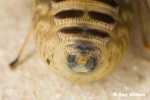
Close-up of a female Tsetse Fly abdomen in a very late stage of pregnancy.
The fly is close to giving birth when the polypneustic lobes at the rear end of the larva turn black and become visible through the female's cuticle.
3. Parturition (birth of the larva)
In the wild, the female Tsetse fly searches for a suitable place to give birth to the larva, usually an area of sheltered, loose, sandy soil. The birthing process is started by massaging the abdomen with the hind legs. The larva is born tail first, with the hard, black polypneustic lobes emerging from the vulva first, preceded by a droplet of clear liquid.
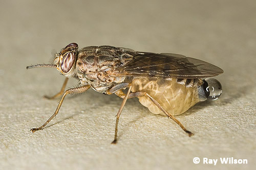
Once the polypneustic lobes are clear of the vulva the rest of the birth proceeds very rapidly and is usually completed in about 5-10 seconds.
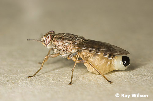
Contractions of the larva's body akin to a backwards crawling movement, help the female expel the larva.
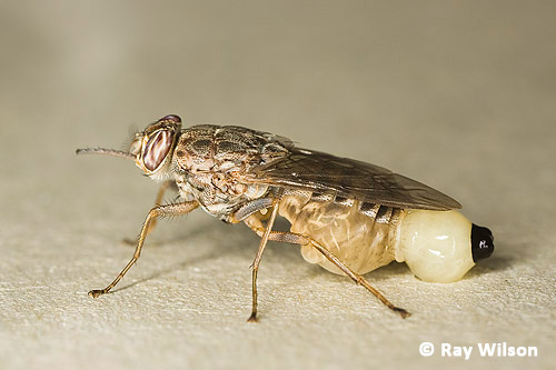
With a final push from the hind legs the larva is free...
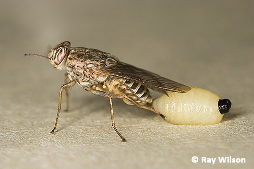
A few seconds after giving birth the female flies away. At 25°C the female Tsetse fly is capable of producing a larva every 7-9 days.
4. Larval & pupal stages
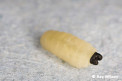
Savannah Tsetse fly (Glossina morsitans morsitans) larva
The newly born larva is fully developed and does not feed outside of the female.
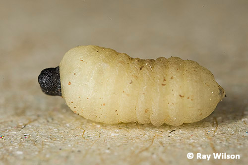
Savannah Tsetse fly (Glossina morsitans morsitans) larva
Immediately after being born, the larva begins to crawls in search of a suitable place to pupate, usually by burrowing underground in the loose, sandy soil. It moves via successive contractions of its body segments that move in a wave down the length of its body starting at the head and working their way back towards the polypneustic lobes.
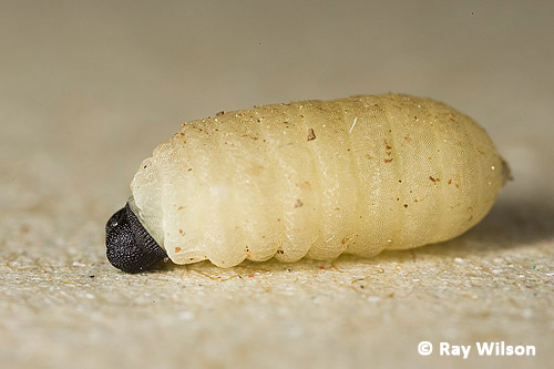
Savannah Tsetse fly (Glossina morsitans morsitans) larva
The larva begins its transformation into a pupa by rounding up into a barrel shape. This process usually starts about 1 hour after parturition.
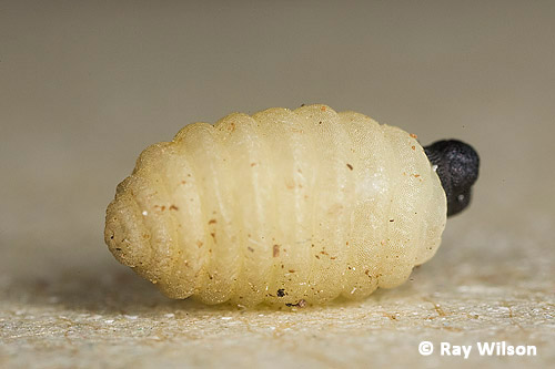
Savannah Tsetse fly (Glossina morsitans morsitans) larva transforming into a pupa
Over the space of a few hours the pupa hardens and darkens until it is a dark purplish-brown colour. The hard outer casing of the pupa is termed the puparium.
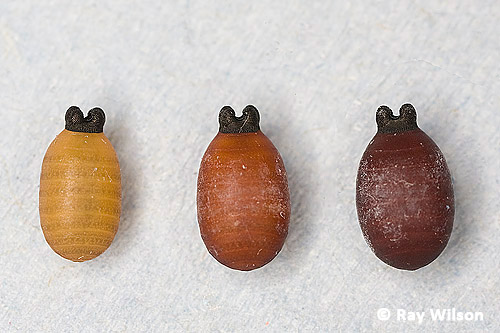
Stages of development of Savannah Tsetse fly (Glossina morsitans morsitans) pupae
5. Eclosion (Emergence from the puparium)
About one month after pupation, the fully developed adult breaks out of the puparium with the aid of an inflatable balloon-like structure on its head called the ptilinum.
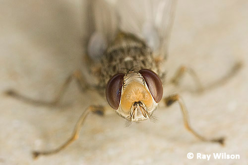
newly eclosed female Savannah Tsetse fly (Glossina morsitans morsitans) with fully extented ptilinum
Once it has escaped the confines of its subterranean pupal case, the soft-bodied fly pushes its way through the soil towards the surface by a combination of thoracic and abdominal contractions (not too dissimilar from the movement of larvae), and, when it encounters resistance from an obstruction, repeatedly inflating the ptilinum in a series of pulses to provide extra force.
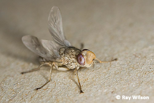
newly eclosed female Savannah Tsetse fly (Glossina morsitans morsitans) with fully extented ptilinum
After it reaches the surface and is able to walk freely, the ptilinum is deflated and the structure recedes back into the head until only a slight crease, called the ptilinal suture, is visible.
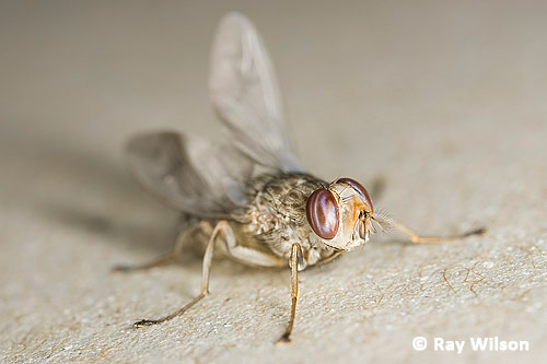
newly eclosed female Savannah Tsetse fly (Glossina morsitans morsitans) with ptilinum receding
This flightless stage of development is sometimes refered to as the "spider" stage due to the fly's tendency to scuttle about like a spider.
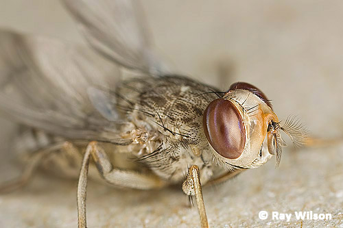
newly eclosed female Savannah Tsetse fly (Glossina morsitans morsitans) with ptilinum receding
At this point the fly begins to inhale air to expand its still soft body to its full size. The difference in size between a freshly eclosed fly and a hardened, fully expanded fly can be up to 90%! In comparison, flies of non-viviparous species, such as Sarcophaga flesh flies, usually only have about a 30% size difference. About an hour after reaching the surface, the expansion and hardening of the body and wings is complete and the fly is ready to fly off in search of its first bloodmeal.

male Savannah Tsetse Fly (Glossina morsitans morsitans) probing skin with his proboscis
The normal product of diuresis is a clear liquid (as can be seen in the fed female at the top of this page), the cloudy, straw-coloured droplet being excreted by the teneral fly below is its meconium. This comprises the remains of the sloughed, degenerating, larval midgut epithelium and is voided from the adult's gut soon after emergence from the puparium.
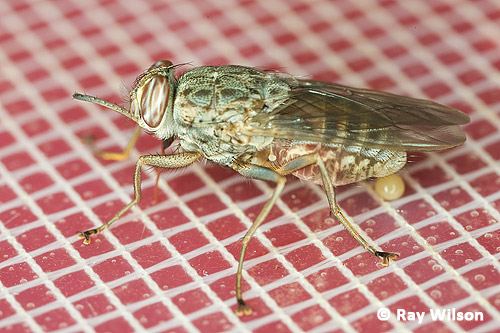
Teneral female Savannah Tsetse fly (Glossina morsitans morsitans) feeding on blood through a silicon membrane and excreting its meconium.
Once the females have fed and mated, the whole cycle begins again.
Feeding Tsetse Flies in the Laboratory
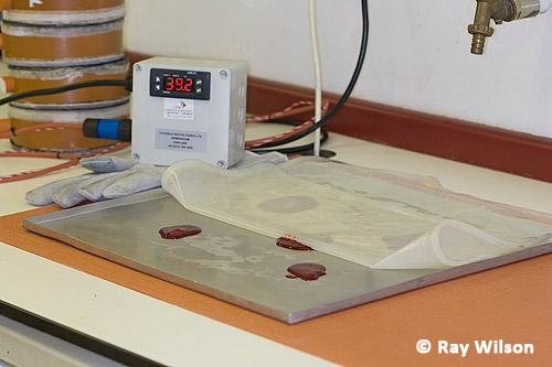
Laboratory feeding apparatus
In the laboratory, routine feeding of the tsetse flies takes place through a silicon membrane. The apparatus used is shown above, and comprises of: a heating mat which is thermostatically controlled to keep the blood at body temperature; a metal tray onto which small quantities of blood are placed; and a silicon membrane which simulates skin for the flies to pierce.
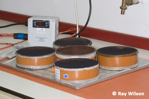
Cages of Tsetse flies being membrane fed
The cages containing the flies are placed on top of the pools of blood and the flies allowed to feed.
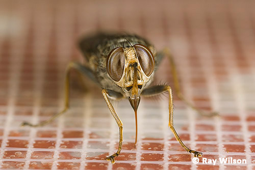
Male SavannahTsetse Fly (Glossina morsitans morsitans) feeding on blood through a silicon membrane
The mouthparts are a useful feature for distinguishing Tsetse Flies from other groups of flies. The proboscis is attached to the head via a bulb-like structure, called the thecal bulb, which contains the muscles necessary for the manipulation of the proboscis. When not feeding, the proboscis is protected by the maxillary palps (the formidable-looking, but actually quite innocuous, saw-like appendage projecting from the fly's head).
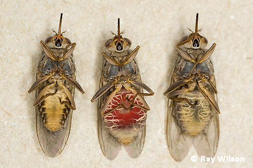
Comparison of female SavannahTsetse Flies (Glossina morsitans morsitans)
left = unfed; middle = immediately after feeding; right = 24 hours after bloodmeal
The abdomen swells enormously after the fly takes a bloodmeal and the extra weight of the blood makes it very difficult for the insect to fly after feeding. In order to regain the power of flight as rapidly as possible, the meal is compacted down to a tight pellet of cells and the excess water excreted. By 24-hours post-bloodmeal, digestion of the blood is well underway and the fly has slimmed down considerably. The blackish colour is due to the breakdown of haemoglobin.
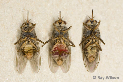
Comparison of male SavannahTsetse Flies (Glossina morsitans morsitans)
left = unfed; middle = immediately after feeding; right = 24 hours after bloodmeal
Males don't generally take such a large meal as the females and retain a slimmer profile throughout the process.
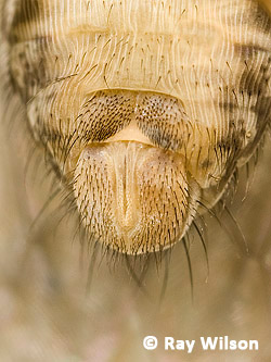 Male |
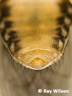 Female |
Comparison of external genetalia of male and female Glossina morsitans morsitans
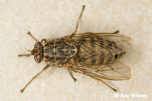
Dorsal view of SavannahTsetse Fly (Glossina morsitans morsitans)
Note the diagnostic "hatchet cell" in the wing venation visible in the above dorsal view. This is formed by a strong curvation of the basal part of vein 4 immediately at the point it meets the anterior cross vein.
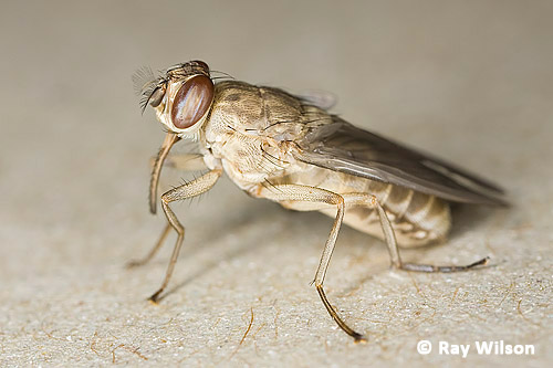
female Riverine Tsetse fly (Glossina palpalis)
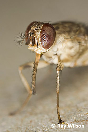
female Riverine Tsetse fly (Glossina palpalis) - the spiky maxillary palps form a protective sheaf for the proboscis when the fly is resting.
All of the Tsetse Flies in these photos were live, uninfected, laboratory reared individuals and photographed under controlled conditions. The comparison photos showing the undersides of the flies were taken in a cold room (4-8°C) to render the flies inactive.
Many thanks to "Flygirl" for all her expert assistance and for bravely allowing the flies to sit on her arm while I photographed them (the bite of the Tsetse fly is painful!).
Ray Wilson owns the copyright of all images on this site.
They may not be used or copied in any form without prior written permission.
raywilsonphotography@googlemail.com
